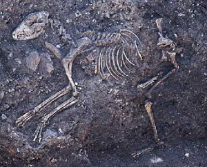
| The dog burial |
| This is more characteristic of the jaws of a modern English Bull Terrier
or Rotweiller. Also the tympanic bullae, the acoustic cavities of the inner
ear, are relatively very large and of a size more compatible with the skull
of a modern greyhound.
The dog exhibits the congenital absence of the lower third molars.This condition is caused by lack of room for the eruption of the rearmost teeth, and occurs because diminution of the jawbone in smaller dogs proceeds faster than the reduction in the size of the teeth. The dog from Silchester is not particularly small, and it is interesting to see the phenomenon in mandibles of this size. It may be that it is an inherited characteristic from a small parent. The muzzle of the dog, although not large, is noticeably unrefined in that there is little variation in the width of the anterior section of the palate. There is little concavity in the frontal area, and the orbits are again of the size seen today in the medium-sized terriers. |
We can calculate the occipital angle of this skull - the angle described
between the basal plane of the cranium and the occipital bone - and this
can suggest the carriage of the head. For example, greyhounds, with
a very low head carriage, have an occipital angle of less than 90o and
this can be as low as 85o, small terriers score between 92o and 95o and
larger terriers, labradors and alsations produce angles between 97o and
120o. This dog from Silchester produces 91o, suggesting that it carried
the head low.
The dog is older than four years; all the bones are fused, and the molar
teeth are very well worn although tooth wear in dogs is notoriously variable.
The right tibia and fibula are fused together, which is a common manifestation
in elderly dogs, and osteomata (button warts) such as that visible on the
skull at the fronto-parietal suture are again frequently seen in old dogs.
|
| The arthropathies observed on the upper vertebrae - periarticular exostosis
on the upper cervical and thoracic bodies - may also be a reflection of
the animal’s maturity. These manifestations are common in all older
animals. However, the more advanced arthropathy in the lumbar vertebrae,
leading to complete fusion of two vertebral bodies in the lower spine (spondylosis),
could be associated with the traumatic injuries noted in the second lumbar
vertebra where the neural spine has been fractured and displaced to the
left. Indeed, injuries to the back of the animal associated with
subsequent infection can be seen in three sections of the thoracic spine
also, that involving the 9th thoracic vertebra showing that the infection
had lodged within the bone of the neural spine to set up a draining osteomyelitis.
It would appear that the vertebral arthropathies may result from injuries
to the neural spines, the most likely cause of these intermittent manifestations
being blows across the back.
Further trauma is visible in the forelimbs in the left ulna and the
left fifth metacarpal, the latter being in an early stage of healing and
infected, so the animal would have been lame at the time of death.
Further slight evidence of infection is in the sequestra and surface pitting
visible on the left radius and humerus, and on the right fourth metacarpal
which again would have caused the animal to limp. Toe bones also
indicate infection or the growth of reactive bone at the joint margins.
|
There are two separate traumatic events visible in the skull.
The first is a healed depressed fracture of the forward part of the muzzle,
and this would have been due a substantial blow. It was sufficient
to realign the nasal bones downwards, causing a malocclusion of the incisor
and canine teeth. This misalignment is very obvious in the the lower
canines, particularly the left, where the “normal” wear on the anterior
edge is visible, as is another “abnormal” plane of wear on the posterior
edge which would not normally be in occlusion. These areas of wear
cannot have happened simultaneously, and the rearrangement of the muzzle
following the trauma almost certainly caused the phenomenon. The
right lower canine has been worn almost to a stump, again indicating malocclusion
and asymmetry of the jaw following the injury. The upper third and
fourth incisors on the left are also missing, and although the tooth sockets
have filled there is clear evidence that infection followed the tooth loss.
The dog suffered another blow to the muzzle after the first injury had fully healed. A transverse fracture is clearly visible some 40 mm from the muzzle tip, and there is evidence of early healing around the fracture margins. The injury was probably not sufficient to kill the dog, but the small degree of healing shows that the animal died shortly after it was sustained. |
|
Silchester
Home | Archaeology
Home | Reading
Home © 2002 Reading University Archaeology Department.
|